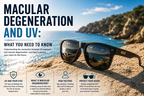

Macular Degeneration and UV: What You Need to Know
Age-related macular degeneration (AMD) is the leading cause of irreversible vision loss in Americans over 50, affecting approximately 11 million people in the United States. Unlike cataracts — which are surgically correctable — AMD causes permanent central vision loss that cannot be restored. UV exposure is a documented contributing risk factor for AMD through oxidative stress mechanisms in the retinal pigment epithelium. Reducing lifetime UV dose is one of the modifiable interventions for AMD risk reduction, alongside smoking cessation and dietary antioxidant intake. For the complete UV eye disease overview, see the complete guide to UV and eye disease.

1. What AMD Is: The Macula and Its Failure
The Macula and Central Vision
The macula is a small, specialized region at the center of the retina — approximately 5mm in diameter — that is responsible for high-acuity central vision. It contains the highest density of cone photoreceptors in the retina, including the fovea at its center, which provides the sharpest visual detail used for reading, face recognition, driving, and any task requiring fine visual discrimination. The macula's optical design — high cone density, specialized photoreceptor types, and dedicated neural processing pathways — makes it the most visually productive region of the retina and the region whose loss has the most severe functional consequences.
AMD causes progressive deterioration of the macula through mechanisms that damage photoreceptors and their supporting infrastructure. Advanced AMD produces a central scotoma — a blind spot in the center of the visual field — that eliminates the detailed vision required for most daily visual tasks. Peripheral vision is typically preserved, allowing mobility and basic orientation, but the reading, face recognition, and driving that depend on central acuity are lost.
Dry vs Wet AMD
AMD has two forms with different progression rates and treatment options:
- Dry (atrophic) AMD: the most common form, accounting for approximately 85–90% of AMD cases. Characterized by the progressive accumulation of drusen (lipid-protein deposits) beneath the retinal pigment epithelium (RPE) and the gradual atrophy of RPE cells and overlying photoreceptors. Dry AMD progresses slowly over years to decades; advanced dry AMD (geographic atrophy) causes significant central vision loss. No approved treatments exist for geographic atrophy as of the early 2020s, though several are in late-stage trials.
- Wet (neovascular) AMD: less common but more rapidly progressive. Characterized by the growth of abnormal new blood vessels (choroidal neovascularization) beneath the retina that leak fluid and blood, causing rapid macular damage and vision loss. Wet AMD can cause severe vision loss within weeks to months without treatment. Anti-VEGF injections (bevacizumab, ranibizumab, aflibercept) can slow or stabilize wet AMD progression in many patients but do not restore lost vision.
2. The UV-AMD Connection: Mechanisms and Evidence
Why the Macula Is Vulnerable to UV
The macula's vulnerability to UV-related damage is paradoxical — UV-B is largely absorbed by the lens before reaching the retina, and UV-A penetration to the macula is limited by the lens. Several factors make the macula disproportionately susceptible to UV-related oxidative stress despite these protective filters:
- High metabolic rate: the macula's dense photoreceptor population and high visual processing activity create an exceptionally high oxygen consumption rate — the highest in the body per unit volume of tissue. High metabolic activity generates reactive oxygen species (ROS) as metabolic byproducts, creating a baseline oxidative stress environment in the macula even without UV exposure.
- Lipofuscin accumulation: the RPE cells that support photoreceptors accumulate lipofuscin — a mixture of oxidized lipids and proteins generated from incomplete digestion of shed photoreceptor outer segments — over decades. Lipofuscin is a potent photosensitizer that generates ROS when exposed to light, including the UV-A and visible blue light that reaches the macula. Lipofuscin accumulation amplifies the oxidative stress from light exposure in the aging macula.
- Polyunsaturated fatty acid concentration: photoreceptor outer segments are rich in polyunsaturated fatty acids (particularly DHA), which are highly susceptible to lipid peroxidation from ROS attack. UV-generated ROS in the macula preferentially attack these lipids, generating lipid peroxidation products that damage adjacent cell structures.
- Limited antioxidant replenishment: like the lens, the macula's antioxidant defense capacity — lutein, zeaxanthin, and enzymatic antioxidants — can be depleted by sustained oxidative stress faster than it is replenished, particularly in aging individuals with reduced systemic antioxidant capacity.
The RPE: The Critical Failure Point
The retinal pigment epithelium (RPE) is the single cell layer that lies directly beneath the photoreceptors and provides essential support functions: recycling visual pigments, phagocytosing shed photoreceptor outer segments, maintaining the photoreceptor microenvironment, and forming part of the blood-retinal barrier. RPE cells do not regenerate when lost — they are post-mitotic and do not divide to replace damaged or dead cells in adults. This non-regenerative nature makes RPE cells the critical failure point in AMD: once RPE cells die, the overlying photoreceptors lose their support infrastructure and eventually degenerate as well.
UV-related oxidative stress in the RPE — from UV-A penetration, from lipofuscin photosensitization, and from the oxidative environment of high metabolic activity — accumulates over decades, progressively damaging RPE cells and reducing their functional capacity. The accumulation of drusen beneath the RPE is a marker of RPE stress — drusen are partly composed of lipids and proteins that RPE cells were unable to process, reflecting compromised RPE function. Advanced drusen accumulation and RPE atrophy are the defining features of geographic atrophy (advanced dry AMD).
Epidemiological Evidence
The AMD-UV association is supported by multiple epidemiological studies, though the relationship is less consistently demonstrated than the well-established cataract-UV association:
- West et al. 1989 (Chesapeake Bay): the same population study that established the UV-cataract connection found that higher sun exposure was associated with increased AMD risk, with a statistically significant dose-response relationship for UV-A specifically. This was among the first studies to suggest a UV component in AMD etiology.
- The Blue Mountains Eye Study: found associations between outdoor sun exposure and AMD prevalence in an Australian population, with the relationship most apparent for geographic atrophy (advanced dry AMD).
- The Rotterdam Study: a large Dutch cohort study found that iris color (a proxy for ocular melanin and UV filtration) was associated with AMD risk — lighter-colored irides (less melanin, more UV penetration) were associated with higher AMD risk, suggesting a UV contribution to AMD etiology.
- EUREYE Study: a pan-European case-control study found that lifetime sunlight exposure was associated with early and late AMD, with the association strongest in those with low antioxidant levels — suggesting that UV and antioxidant deficiency interact in AMD risk.
Why the Evidence Is Less Definitive Than for Cataracts
Several factors make the AMD-UV association less clear-cut than the cataract-UV association:
- AMD is more genetically determined than cataracts — complement factor H (CFH) and ARMS2 gene variants account for a large proportion of AMD heritability, making environmental factors (including UV) relatively smaller contributors to total population risk.
- UV penetration to the macula is limited by lens absorption — the macular UV dose is much lower than the lens UV dose, making the pathway to macular damage more indirect.
- AMD's multifactorial etiology (smoking, cardiovascular disease, genetics, diet) makes UV's specific contribution difficult to isolate in population studies.
- Long latency (40–60 years between UV accumulation and clinical AMD) makes accurate UV exposure reconstruction in retrospective studies difficult.
Despite these complexities, the biological mechanisms (lipofuscin photosensitization, RPE oxidative stress, RPE non-regenerative nature) and the available epidemiological associations support UV exposure as a meaningful contributor to AMD risk — one that UV400 protection can address.
3. AMD Risk Factors: UV in Context
| Risk Factor | Strength of Evidence | Modifiable? | Intervention |
|---|---|---|---|
| Age | Very strong — strongest risk factor | No | None |
| Genetics (CFH, ARMS2 variants) | Very strong — 50%+ of risk attributable | No | Genetic testing for risk stratification |
| Smoking | Strong — 2–4x elevated risk in smokers | Yes | Smoking cessation |
| Cardiovascular disease / hypertension | Moderate | Partially | Cardiovascular risk management |
| UV exposure | Moderate — supported by mechanism and epidemiology | Yes | UV400 sunglasses |
| Diet (low antioxidants) | Moderate | Yes | AREDS2 supplements; leafy greens |
| Obesity | Moderate | Yes | Weight management |
| Light iris color | Moderate — proxy for less melanin UV filtration | No | UV protection particularly important |
4. AMD Treatment: Why Prevention Matters More Than for Cataracts
Dry AMD: No Proven Treatment for Geographic Atrophy
For dry AMD before the geographic atrophy stage, AREDS2 supplementation (vitamin C, vitamin E, lutein, zeaxanthin, zinc) reduces the risk of progression to advanced AMD in patients with intermediate AMD by approximately 25%. This is meaningful but partial — it reduces progression risk, not disease reversal. For geographic atrophy (advanced dry AMD), no treatment has been proven to restore lost vision. Several complement inhibitor therapies have received approval or are in late-stage trials, with modest effects on slowing geographic atrophy progression — not restoration.
Wet AMD: Treatable But Not Reversible
Anti-VEGF injections for wet AMD are among the most significant advances in ophthalmology of the past two decades — they can stabilize vision and even produce modest improvement in some patients by suppressing the abnormal blood vessel growth that causes rapid macular damage. However, wet AMD treatment requires repeated injections (monthly to quarterly) indefinitely, is not effective for all patients, and produces vision improvement in only a minority — most patients achieve stabilization rather than improvement. Lost macular photoreceptors are not restored by any current treatment.
The Prevention Imperative
The irreversibility of AMD photoreceptor loss makes prevention substantially more valuable for AMD than for cataracts. A cataract that is not prevented will require surgery — an effective intervention with good visual outcomes. AMD that is not prevented may progress to geographic atrophy or wet AMD causing permanent central vision loss with no effective restoration. The asymmetry between AMD prevention (potentially effective) and AMD treatment (partially effective at best) makes the prevention case for AMD stronger than for any other UV-related eye condition.
5. Lutein, Zeaxanthin, and Macular Pigment
The Macular Pigment as a UV Filter
The macula contains high concentrations of lutein and zeaxanthin — carotenoid pigments that give the macula its characteristic yellow color (macula lutea means "yellow spot"). These pigments function as a partial light filter in the macula, absorbing blue light and some UV wavelengths before they reach the photoreceptors and RPE. Macular pigment optical density (MPOD) is measurable and varies between individuals — higher MPOD is associated with reduced oxidative stress in the macula and lower AMD risk in some studies.
Dietary and Supplemental Sources
Lutein and zeaxanthin are obtained from diet — dark leafy greens (kale, spinach, collard greens), eggs, and corn are primary dietary sources. AREDS2 supplementation (10mg lutein / 2mg zeaxanthin daily) was associated with reduced AMD progression risk in the AREDS2 clinical trial. The combination of dietary lutein and zeaxanthin with UV400 sunglasses addresses AMD risk through complementary mechanisms — internal macular pigment filtration plus external UV blocking.
6. Practical UV Protection for AMD Prevention
Who Should Prioritize AMD Prevention Through UV Protection
- Anyone over 50 with a family history of AMD — genetic risk combined with UV accumulation elevates total risk
- Light-eyed individuals (blue, green, gray irises) — less natural UV filtration through the iris
- Current and former smokers — smoking and UV are additive AMD risk factors; stopping smoking and adding UV protection addresses two modifiable factors simultaneously
- Outdoor workers and people with high lifetime UV accumulation — elevated UV dose makes UV reduction more impactful
- Individuals who have already been diagnosed with early or intermediate AMD — slowing progression through UV reduction and AREDS2 supplementation
Lens Specification for AMD Prevention
UV400 polycarbonate lenses block 100% of UV-A and UV-B — both wavelengths that contribute to macular oxidative stress through direct penetration and lipofuscin photosensitization. Browse UV400 polarized polycarbonate options at navieyewear.com. For individuals particularly concerned about AMD and blue light exposure from screens, blue-light-filtering lenses are available — though the evidence for blue light's contribution to AMD is less established than for UV.
Frequently Asked Questions
Can UV cause macular degeneration?
UV exposure is a documented contributing risk factor for AMD — supported by epidemiological associations and biological mechanisms involving oxidative stress in the retinal pigment epithelium. UV is not the primary or sole cause of AMD (genetics and aging are the dominant factors), but it contributes to the oxidative stress burden in the macula that accumulates over decades toward clinical disease. UV400 protection reduces this contribution.
Is AMD reversible?
No — photoreceptor and RPE cell loss in AMD is irreversible with current treatments. Anti-VEGF injections for wet AMD can stabilize or modestly improve vision by suppressing abnormal blood vessel growth, but do not restore lost photoreceptors. Geographic atrophy (advanced dry AMD) has no proven treatment for restoration. The irreversibility of AMD makes prevention substantially more valuable than treatment — consistent UV400 protection addresses a modifiable contributing risk factor for a disease that cannot be effectively reversed.
What is the best way to prevent macular degeneration?
No intervention guarantees AMD prevention, but the evidence-supported modifiable risk factor interventions are: smoking cessation (the strongest modifiable risk factor), UV400 sunglasses for cumulative UV dose reduction, AREDS2 supplementation for individuals with intermediate AMD, dietary lutein and zeaxanthin from dark leafy greens, cardiovascular risk management, and healthy body weight. These interventions address different AMD risk pathways and are complementary — the strongest risk reduction comes from addressing multiple modifiable factors simultaneously.
Do sunglasses help prevent AMD?
UV400 sunglasses reduce the UV-driven oxidative stress component of AMD risk by blocking the UV wavelengths that contribute to RPE oxidative damage through direct absorption and lipofuscin photosensitization. The magnitude of risk reduction from UV protection alone is not precisely quantified, but the biological mechanism is well-established and the epidemiological associations support UV as a meaningful AMD risk contributor. UV protection is the recommended component of AMD prevention alongside smoking cessation and dietary measures.
How does AMD affect daily life?
AMD affects central vision — the vision used for reading, face recognition, driving, and detailed visual tasks. Peripheral vision is typically preserved, allowing orientation and mobility, but the functional losses from central vision impairment are severe: inability to read standard print, inability to recognize faces, loss of driving ability, and significant limitations in all activities requiring fine visual detail. Advanced AMD is a major cause of loss of independence in older adults — the prevention case addresses not just visual acuity but functional independence.
At what age should I start protecting my eyes for AMD prevention?
As early as possible — the cumulative dose model means that UV reduction at any age reduces the lifetime UV contribution to macular oxidative stress. The highest-return period for UV protection is early adulthood, when UV accumulation rates are highest and the long latency period means protection now prevents disease in 30–40 years. AMD prevention through UV protection is a lifelong strategy, not an intervention begun at 50 when symptoms might first appear.
The Bottom Line
AMD is the leading cause of irreversible central vision loss in Americans over 50, and it cannot be effectively reversed with current treatments. UV exposure contributes to the macular oxidative stress that drives RPE cell damage and photoreceptor loss over decades. UV400 sunglasses reduce this UV contribution — one of the few modifiable AMD risk factors available. Given AMD's irreversibility, prevention through UV reduction, smoking cessation, and dietary antioxidant intake is the most effective AMD management strategy available. Consistent UV400 polarized protection from early adulthood through the senior years reduces the UV-driven component of AMD risk across the decades that matter. navieyewear.com/collections/polarized — Buy 1, Get 3 Free for $119.
Related Reading
- UV and Eye Disease: The Complete Guide | Navi Eyewear
- UV and Cataracts: Prevention, Risk Factors, and Research | Navi Eyewear
- Best Sunglasses for Men Over 50 | Navi Eyewear
- Best Sunglasses for Women Over 50 | Navi Eyewear
- UV Protection for Children: The Complete Family Eye Health Guide | Navi Eyewear
- Sunglasses by Season: The Complete Year-Round UV Guide | Navi Eyewear
- Sunglasses for Outdoor Workers | Navi Eyewear
- Shop Polarized UV400 Sunglasses | Navi Eyewear
Sources & Citations
[1] West SK, et al. "Exposure to sunlight and other risk factors for age-related macular degeneration." Archives of Ophthalmology, 1989. View source →
[2] Cruickshanks KJ, et al. "Sunlight and the 5-year incidence of early age-related maculopathy." Archives of Ophthalmology, 2001. View source →
[3] Fletcher AE, et al. "Sunlight exposure, antioxidants, and age-related macular degeneration." Archives of Ophthalmology, 2008. View source →
[4] AREDS2 Research Group. "Lutein/zeaxanthin for the treatment of age-related macular degeneration." JAMA Ophthalmology, 2013. View source →
[5] Sliney DH. "Ocular exposure to environmental light and ultraviolet." Journal of AAPOS, 2014. View source →
[6] WHO. "Global solar UV index: a practical guide." World Health Organization, 2002. View source →
[7] Chua J, et al. "Genetic and non-genetic risk factors for age-related macular degeneration." Clinical Genetics, 2019. View source →